Cone Beam CT Scan
Cone-beam computed tomography systems (CBCT) are a variation of traditional computed tomography (CT) systems. The CBCT systems used by dental professionals rotate around the patient, capturing data using a cone-shaped X-ray beam. These data are used to reconstruct a three-dimensional (3D) image of the following regions of the patient’s anatomy: dental (teeth); oral and maxillofacial region (mouth, jaw, and neck); and ears, nose, and throat (“ENT”).
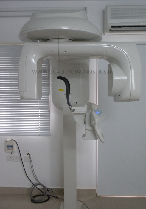
First Cone Beam CT Scan (CBCT) in Chennai
Quality Diagnostics, the distinction of acquiring south India’s very first CBCT (Cone Beam Computed Tomography) unit in 2010
Advantages of Cone Beam CT Scan
X-ray imaging, including dental CBCT, provides a fast, non-invasive way of answering a number of clinical questions. Dental CBCT images provide three-dimensional (3-D) information, rather than the two-dimensional (2-D) information provided by a conventional X-ray image. This may help with the diagnosis, treatment planning and evaluation of certain conditions.
Although the radiation doses from dental CBCT exams are generally lower than other CT exams, dental CBCT exams typically deliver more radiation than conventional dental X-ray exams. Concerns about radiation exposure are greater for younger patients because they are more sensitive to radiation (i.e., estimates of their lifetime risk for cancer incidence and mortality per unit dose of ionizing radiation are higher) and they have a longer lifetime for ill-effects to develop.